Scientific illustration
Scientific illustration plays a key role in specimen-based paleobiological research. Anatomically accurate reconstructions of extinct organisms serve two main purposes. First, illustrations allow us to graphically summarize the morphological information preserved in body fossils, and help us to visualize extinct organisms as actual living beings. Second, illustrations represent an excellent resource for teaching and outreach. It is much easier to communicate the biological complexity of an extinct animal to a general audience through an aesthetically attractive reconstruction, than by solely examining the fossil data, as the latter is often incomplete or distorted by preservation biases. This section showcases a combination of schematic diagrams, artistic impressions, and professional scientific reconstructions that accompany our published manuscripts.
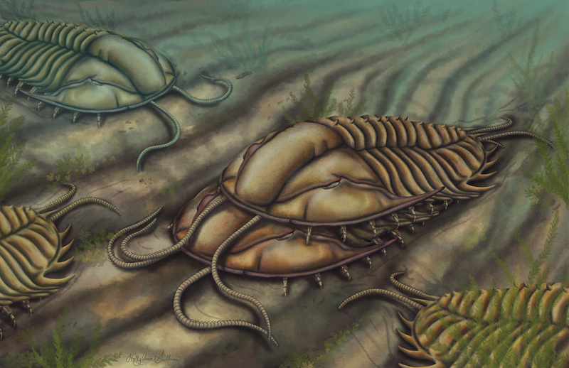
Artistic reconstruction of the Burgess Shale (mid-Cambrian) trilobite Olenoides serratus during amplex, in which the male (top) grasps the pygidial spines of the female (bottom) using specialized clasper-like appendages (see publication here). Artwork by Holly Sullivan.

Left: Artistic reconstruction of the middle Cambrian comb jellies Ctenorhabdotus campanelliformis (top) and Thalassostaphylos elegans (bottom) from the Marjum Formation in Utah (see publication here). Artwork by Holly Sullivan.
Right: Sketch for artistic reconstruction of Ctenorhabdotus campanelliformis in lateral (top) and apical (bottom) views. Artwork by Holly Sullivan.

Top: Artistic reconstruction of the Cambrian (Drumian) Marjum biota in the House Range of Utah, USA (see publication here). Artwork by Holly Sullivan. Number key for taxa illustrated: 1. Scathascolex minor?; 2. Diagoniella cyathiformis; 3. Hyolithes sp.; 4. Modocia typicalis; 5. Marpolia-like alga; 6. Leptomitella metta; 7. Peytoia nathorsti; 8. Pahvantia hastata; 9. Cubozoan jellyfish; 10. Perspicaris? ellipsopelta; 11. Oesia disjuncta/Margaretia dorus; 12. Tuzoia guntheri; 13. Bathyuriscus fimbriatus; 14. Sphenoecium wheelerensis; 15. Canthylotreta marjumensis; 16. Castericystis vali; 17. Choia hindei; 18. Caryosyntrips camurus?; 19. Branchiocaris pretiosa?; 20. Gogia spiralis; 21. Buccaspinea cooperi; 22. Itagnostus interstrictus; 23. Chancelloria sp. |
Bottom: Sketch for environmental reconstruction. Artwork by Holly Sullivan. |

Top: Environmental reconstruction of the early Cambrian (Stage 3) Chengjiang biota in South China, featuring the eudemersal radiodont Cambroraster sp. (see publication here). Artwork by Holly Sullivan. |
Bottom Left: Sketch for Cambroraster sp. reconstruction. Artwork by Holly Sullivan. |
Bottom Right: Sketch for environmental reconstruction. Artwork by Holly Sullivan. |